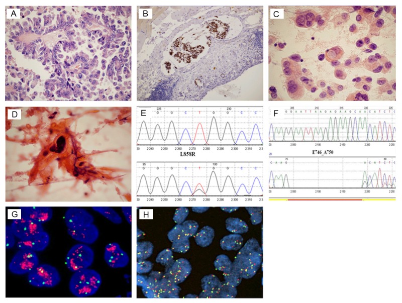
Figure 1.

Images of the most frequent histological and molecular results [A: Adenocarcinoma, papillary subtype (HE, 400 x), B: Embolism of adenocarcinoma cells in the lymphatic vessel (TTF-1, 100 x), C: Adenocarcinoma with visible droplets of intracytoplasmic mucus (HE, 400 x), D: Squamous cell carcinoma (HE, 1000 x), E, F: Graphical illustration of EGFR gene mutations: exon 21 substitution (L858R) and exon 19 deletion (E746_A750) respectively, G, H: FISH positive results: amplification (1000 x) and high polysomy (600 x)].
