
Figure 2.
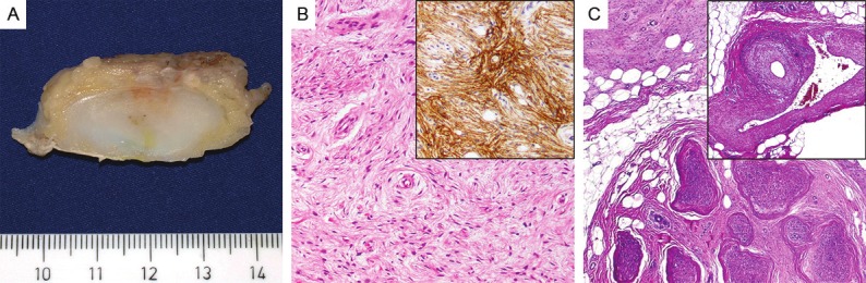
The resected tumor had ill-defined margins and a pale fleshy, solid cut-surface (A). On microscopic view, the tumor cells were arranged in parallel oriented fascicles with a focally whorled growth pattern and showed ovoid to elongated slender nuclei and pale cytoplasm (B, H&E, x100) with expression of EMA (B, inset). Perineurial proliferations were seen extending from the perineurium of the sural nerve into surrounding adipose tissue (C, H&E, x40), Striking vasculopathy was seen within surrounding fat with prominent thickening of arterial and venous vessels (C, inset, H&E, x40).