Figure 2.
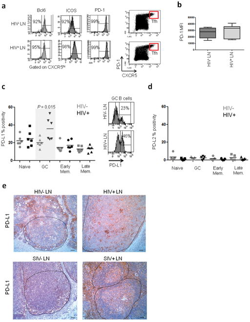
Ex-vivo characterization of Tfh cells and B cells in LNs from HIV-infected and uninfected individuals. (a) Enrichment of Tfh cells in the CXCR5hi population of both HIV− and HIV+ LNMCs as determined by Bcl-6, ICOS and PD-1 staining. (b) Expression levels of PD-1 on Tfh cells from HIV− and HIV+ LNMCs as measured by mean fluorescence intensity (MFI). (c) Frequency of PD-L1 expression on B cell subsets from infected and uninfected LNMCs. Subsets were defined as naïve (CD3−CD19+CD38−IgD+), GC (CD3−CD19+CD38hiIgD−), early memory (CD3−CD19+CD38+IgD−) and late memory (CD3−CD19+CD38−IgD−) (HIV− n=6; HIV+ n=6). (d) Frequency of PD-L2 expression on B cell subsets (HIV− n=5; HIV+ n=5). (e) Representative images for PD-L1 staining on LN sections from HIV-uninfected (n=6) and infected (n=5) subjects as well as SIV-uninfected (n=4) and infected (n=2) macaques. Scale bar, 50 μm.

