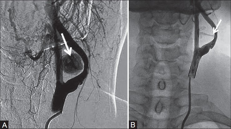
Figure 5(A, B):

Digital subtraction angiography of left internal carotid artery showing the (A) pseudoaneurysm medial to ICA filling up with contrast with narrowed and irregular lumen of ICA (arrow) and (B) narrow neck of the pseudoaneurysm with contrast jet (arrow)
