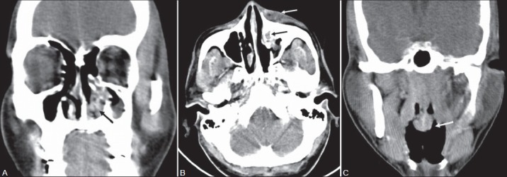
Figure 2(A-C):

A 34-year-old male with left nasal mass and periorbital swelling. Contrast-enhanced CT PNS (A) Coronal image shows an irregular enhancing soft tissue lesion in the left inferior nasal cavity (black arrow) with partial erosion of the left inferior turbinate. (B) Axial CT image shows extension of enhancing soft tissue into the left nasolacrimal duct (black arrow) with left lacrimal sac pyocele (white arrow). (C) Coronal section shows another pedunculated polyploidal lesion (white arrow) with bulbous tip arising from the posterior nasopharyngeal wall. Diagnosis after surgery confirmed as rhinosporidiosis
