Figure 2.

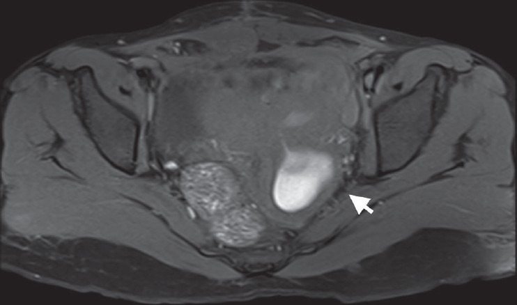
Axial fat-suppressed T1W MRI shows hyperintense contents of dilated left hemivagina (arrow) and uterine cavity, suggestive of blood products

Axial fat-suppressed T1W MRI shows hyperintense contents of dilated left hemivagina (arrow) and uterine cavity, suggestive of blood products