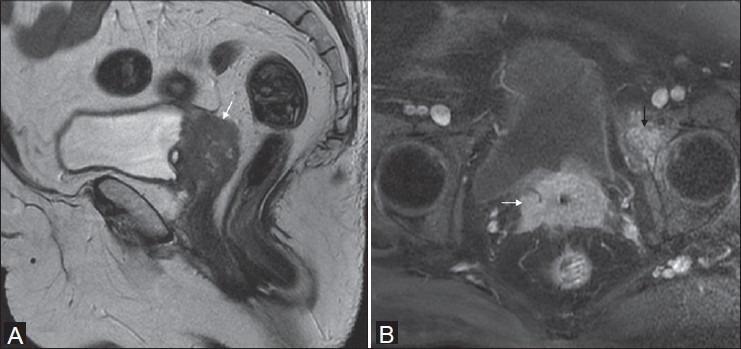
Figure 12(A, B).

Operated case of Ca cervix in a 71-year-old female. Sagittal T2W image (A) shows intermediate signal intensity recurrent mass at vaginal vault (white arrow). Oblique axial T1 fat-suppressed post-gadolinium image (B) shows homogeneous enhancement of the mass (white arrow) with secondary deposit in the anterior column of left acetabulum (black arrow)
