
Figure 6.
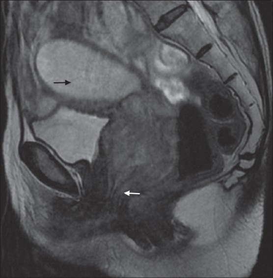
A 65-year-old female with poorly differentiated squamous cell carcinoma (stage III A). Hyperintense mass infiltrating the vaginal fornices and extending caudally to lower third of the vagina along the the anterior and posterior vaginal walls (white arrow). Collection in endometrial cavity is seen as hyperintensity (black arrow)