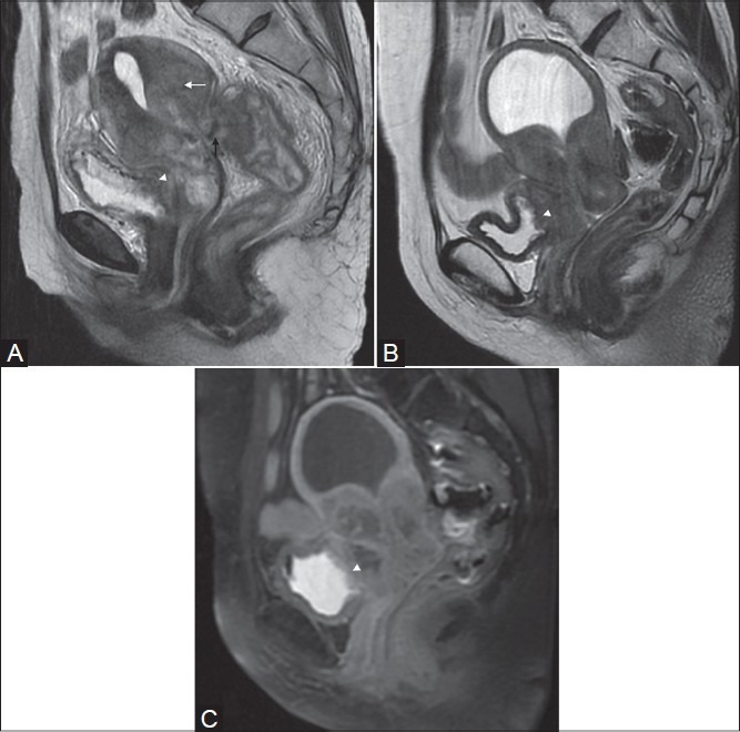
Figure 8(A-C):

Squamous cell carcinoma in two different patients (stage IVA). Sagittal T2W image shows a large mass arising from the cervix and involving the uterine myometrium (white arrow in A) with invasion in the rectum demonstrated as loss of T2-low signal intensity rectal wall (black arrow in A). Also note the infiltration in posterior bladder wall (white arrow-head in A), better seen in the second patient on T2 and post-gadolinium image (white arrow heads in B and C)
