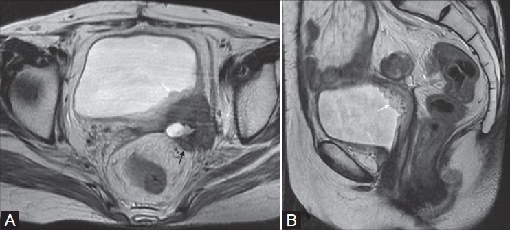
Figure 9(A, B).

Poorly differentiated adenocarcinoma in a 54-year-old lady. Recurrent mass infiltrating left posterolateral bladder wall with hyperintense thickening of the bladder mucosa on T2W images, typically called bulbous edema sign (white arrows in A and B). Note the infiltration of mesorectal fascia and extension in the mesorectum (black arrow in A)
