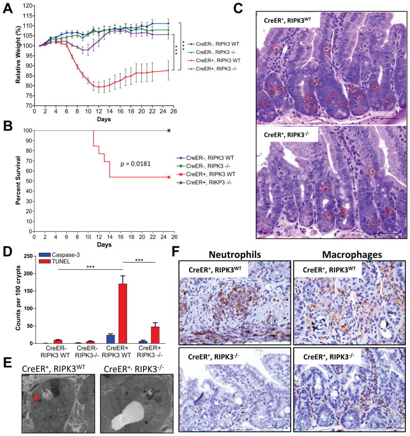
Figure 2. RIPK3 deficiency protects from acute deletion of caspase-8.
(A–F) Rosa26.CreER−, casp8f/f, ripk3+/+ (CreER−, RIPK3WT), Rosa26.CreER−, casp8f/f ripk3−/− (CreER−, RIPK3−/−), Rosa26.CreER+, caspase-8flox/flox, RIPK3+/+ (CreER+, RIPK3WT) and Rosa26.CreER+, casp8f/f, ripk3−/− (CreER+, RIPK3−/−) animals were gavaged with 1mg tamoxifen per 25g animal body weight for 6 consecutive s. Animals were observed over 25 days for weight loss (A) and lethality (B). (C) Day +6 proximal small intestine sections from tamoxifen-treated CreER+, RIPK3WT and CreER+, RIPK3−/− animals were stained with hematoxylin and eosin. Red circles highlight dead cells. (D) Proximal small intestine sections were stained for TUNEL and cleaved caspase-3 and the number of TUNEL-positive or cleaved caspase-3 positive cells per 100 crypts was determined by manual counts. (E) Transmission electron microscopy of proximal small intestine crypts at day +6. Arrow points to necrotic cell. (F) Day +9 proximal small intestine sections from tamoxifen-treated CreER+, RIPK3WT and CreER+, RIPK3−/− animals were immunostained for neutrophils (anti-LY-6B.2) (left panel) or macrophages (anti-F4/80) (right panel). (***)p<0.001.

