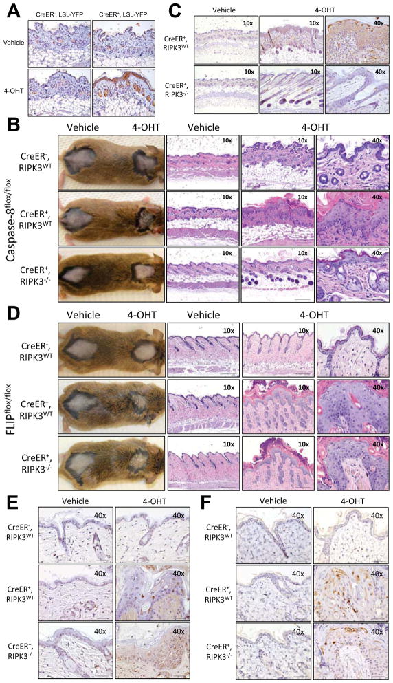
Figure 3. Acute deletion of caspase-8 or FLIP in the skin by 4-hydroxytamoxifen painting produces local inflammation and tissue damage.
Animals were shaved in two distinct dorsal areas. The shaved area around the neck was painted with 4-hydroxytamoxifen (40HT) while the shaved area near the tailbase was painted only with vehicle. (A) Histological sections from the painted skin areas from Rosa26.CreER−, LSL-YFP (CreER−, LSL-YFP) and Rosa26.CreER+, LSL-YFP (CreER+, LSL-YFP) were taken the day after the last painting and stained with anti-GFP antibodies to detect YFP expression. (B–C) Rosa26.CreER−, casp8f/f ripk3+/+ (CreER−, RIPK3WT), Rosa26.CreER+, casp8f/f, ripk3+/+ (CreER+, RIPK3WT) and Rosa26.CreER+, casp8f/f ripk3−/− (CreER+, RIPK3−/−) were painted four times and at day +12 (B) photos and H&E-stained sections or (C) TUNEL-stained sections were produced. (D–F) Rosa26.CreER−, cflarf/f ripk3+/+ (CreER+, RIPK3WT), Rosa26.CreER+, cflarf/f, ripk3+/+ (CreER+, RIPK3WT) and Rosa26.CreER+, cflarf/f, ripk3−/− (CreER+, RIPK3−/−) were painted two times and at day +10 (D) photos and H&E-stained sections, (E) TUNEL-stained sections or (F) cleaved caspase-3 were produced.

