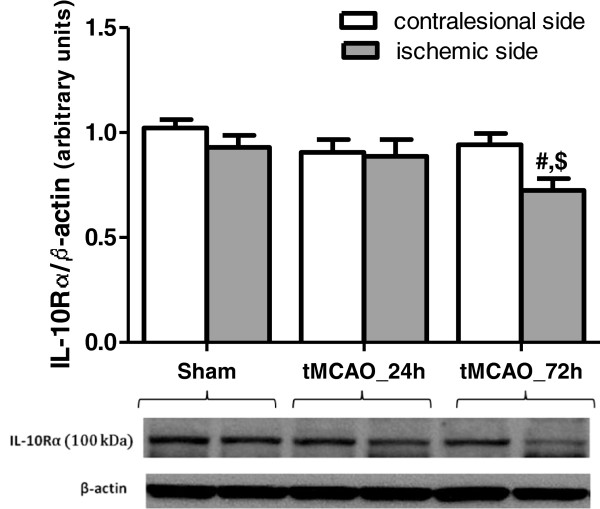
Figure 3.
Temporal IL-10R expression in Wistar rat brains after tMCAO. Western blotting analysis showed no change in IL-10Rɑ protein expression in Wistars brains at 24 after tMCAO but it declined significantly at 72 h relative to baseline. # significantly different from the sham right hemisphere (p < 0.05), $ significantly different from the 72 h time point contralesional hemisphere (p < 0.05), N = 4-7 per group.

