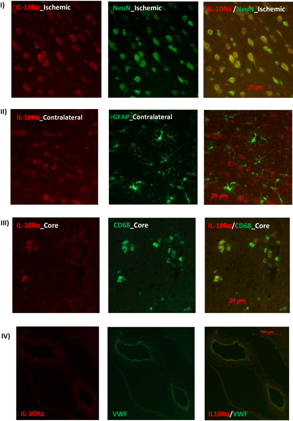
Figure 4.
Immunofluorescent localization of IL-10R in Wistar rat brains after tMCAO. IL-10Rɑ strongly expressed in neurons (NeuN) and endothelial cells (VWF) of sham and stroked animals in addition to activated microglia/macrophages (CD68) after tMCAO but not astrocytes (GFAP). Images were taken from 24 h brain sections. 72 h brain sections showed similar results. N = 4 per group.

