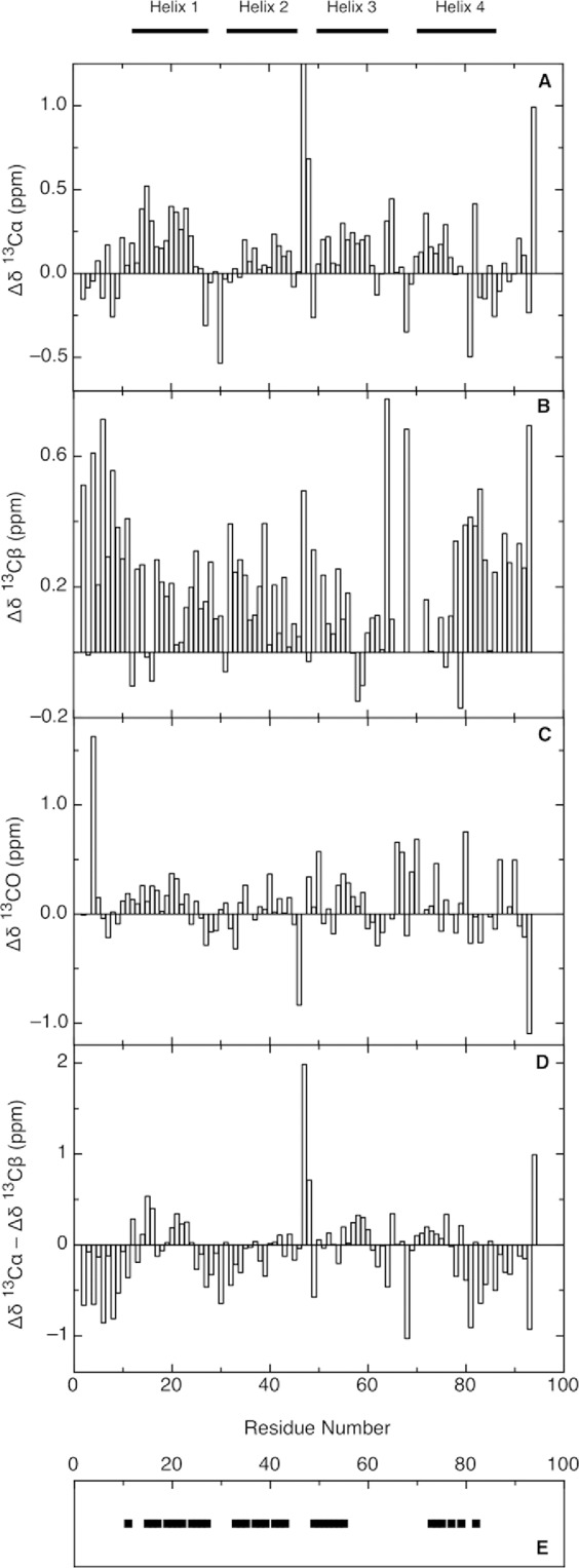
Figure 6.

Chemical shift deviations of Im7H3M3 in the urea-unfolded state (6M urea) for 13Cα (A), 13Cβ (B), and 13CO (C), in 50 mM phosphate buffer, pH 7 at 25°C. Intrinsic random coil chemical shifts were used according to reference.41,42 Secondary chemical shift differences Δδ13Cα − Δδ13Cβ (D) provide a valuable probe of secondary structure propensities; positive values indicate α-structure propensity and negative values indicate β-structure propensity. His 47 show a large 13Cα deviation ∼2.5 ppm (A), nevertheless a linear analysis of chemical shifts (LACS) was performed validating all Im7H3M3 chemical shifts. Summary of the assigned cross-peaks in the 1H-1H-15N NOESY-HSQC spectrum of Im7H3M3 (E) showing NOEs between the HN of residue i and the HN of residue i + 1 (black squares). Though some NOE connectivities between NH resonances could be observed in the three-dimensional NOESY-HSQC spectrum, the severe overlap of the amide resonances limited the number of NOE peaks that could be assigned. The horizontal bars on the top of the figure indicate the secondary structure elements present in the native state.
