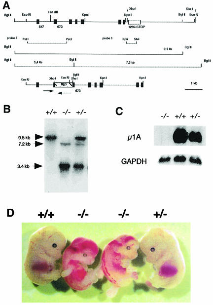
Fig. 1. Targeted disruption of the µ1A-adaptin gene. (A) Genomic 9.5 kb EcoRI µ1A locus (top) and 5.6 kb EcoRI–KpnI targeting construct (bottom). Exons are indicated by black bars. Arrows indicate µ1A and neoR ORF. Lines mark chromosomal DNA fragments generated by BglII digest in control and mutant cells used to demonstrate homologous recombination and the DNA fragments used as internal (probe 1) and external (probe 2) probes for hybridization of BglII-digested chromosomal DNA. (B) Southern blot analysis of BglII-digested chromosomal DNA isolated from amnion epithelia of day 12.5 p.c. embryos hybridized with probe 2. (C) Northern blot of total embryonic fibroblast RNA hybridized first with 32P-labeled mouse µ1A cDNA and then with GAPDH cDNA. (D) Embryos from a litter isolated at day 13.5 of embryonic development.

An official website of the United States government
Here's how you know
Official websites use .gov
A
.gov website belongs to an official
government organization in the United States.
Secure .gov websites use HTTPS
A lock (
) or https:// means you've safely
connected to the .gov website. Share sensitive
information only on official, secure websites.
