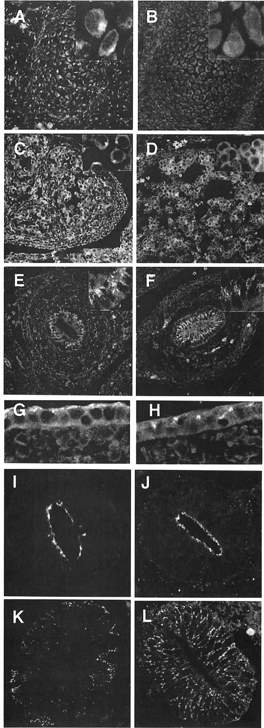
Fig. 5. Labeling of cryosections of the embryos for γ-adaptin staining. Shown are vertebral bodies of control (A) and µ1A-deficient (B) embryos, liver of control (C) and µ1A-deficient (D) embryos, the gut of control (E) and µ1A-deficient (F) embryos, and epidermis of ct (G) and µ1A-deficient (H) embryos. Polarization of gut epithelial cells demonstrated by staining for tight junctions with anti-ZO-1 in ct (I) and µ1A-deficient cells (J) and for the LDL receptor by fluorescent LDL in ct (K) and µ1A-deficient cells (L).

An official website of the United States government
Here's how you know
Official websites use .gov
A
.gov website belongs to an official
government organization in the United States.
Secure .gov websites use HTTPS
A lock (
) or https:// means you've safely
connected to the .gov website. Share sensitive
information only on official, secure websites.
