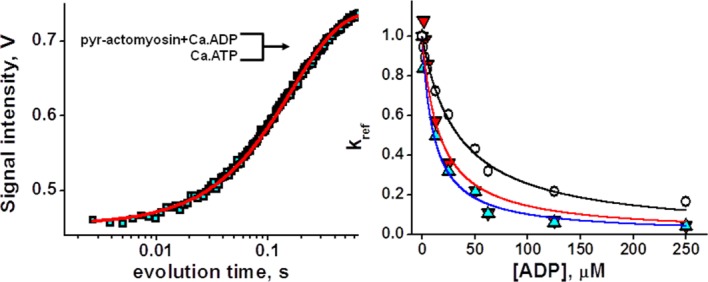
Figure 4.

ATP induced actomyosin dissociation in the presence of ADP. Left, pyrene fluorescence transient after rapid mixing of 50 µM Ca.ATP and 0.15 µM pyrene-actomyosin, premixed, and incubated with 50 µM Ca.ADP. Fit with one exponent. Right, normalized rates of ATP induced actomyosin dissociation in the presence of ADP. Magnesium, upturned triangles, manganese, triangles, calcium, circles, lines, fit. Ca.ADP has weaker affinity to actomyosin, compared with Mg.ADP and Mn.ADP. [Color figure can be viewed in the online issue, which is available at http://wileyonlinelibrary.com.]
