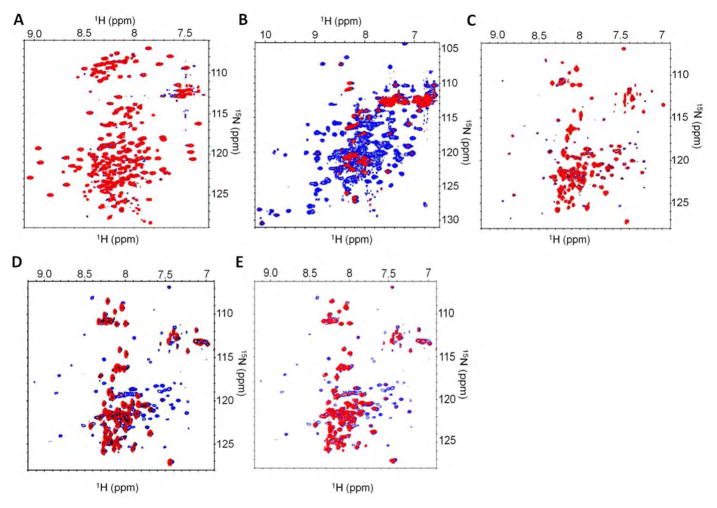
Fig. 7.
PPM1A binds to the C-terminal domain of MAN1. 1H-15N 2D NMR spectra were recorded on 1:0 and 1:1 ratio samples of a 15N-labeled protein and its unlabelled partner. (A) Representative HSQC spectrum of 15N-labeled fragment of MAN1 from amino acid 1 to amino acid 471 (MAN1N) with (red) or without (blue) added PPM1A. N=2 independently generated replicates. (B) Representative HSQC spectrum of 15N-labeled fragment of MAN1 from amino acid 658 to amino acid 911 (MAN1C) with (red) or without (blue) added PPM1A. (C) Representative TROSY spectrum of 15N-labeled PPM1A with (red) or without (blue) addition of the MAN1 fragment from amino acid 1 to amino acid 471 (MAN1N). N=2 independently generated replicates. (D) Representative TROSY spectrum of 15N-labeled PPM1A with (red) or without (blue) addition of the MAN1 fragment from amino acid 658 to amino acid 911 (MAN1C). N=2 independently generated replicates. (E) Representative TROSY spectrum of 15N-labeled PPM1A with (red) or without (blue) addition of the MAN1 fragment from amino acid 755 to amino acid 911 (MAN1Luhm). N=2 independently generated replicates.

