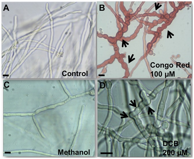
Figure 2. DCB and CR cause the formation of aberrant hyphal structures.
Bright field microscopy pictures (40x in A, B and C, and 60x in D) of mycelium grown in liquid MM (A), in medium supplemented with 100 µM CR (B), 1% v/v MetOH (C) and 200 µM DCB (D). The arrows point to aberrant structures present along the hyphae (namely swollen regions and bulges). Scale bar refers to 10 µm.

