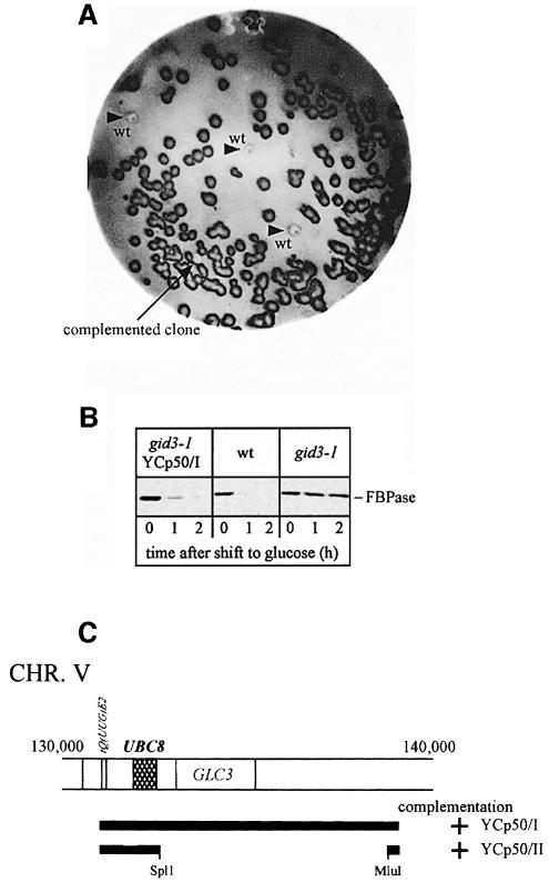
Fig. 1. (A) Colony screen for the detection of gid3-1-complementing genomic DNA fragments. Cells were transferred on nitrocellulose sheets as described in Materials and methods and probed with antibodies against FBPase. Further details are given in the text. gid mutant cells appear significantly darker than wild-type cells (arrowheads) due to their higher FBPase level. (B) Complementation of gid3-1 cells was verified in immunoblots following FBPase immunogenic material after retransformation of the plasmid, isolated from the clone shown in (A). The complementing plasmid re-introduces the FBPase degradation phenotype. (C) Subcloning identifies UBC8 as the complementing ORF.

An official website of the United States government
Here's how you know
Official websites use .gov
A
.gov website belongs to an official
government organization in the United States.
Secure .gov websites use HTTPS
A lock (
) or https:// means you've safely
connected to the .gov website. Share sensitive
information only on official, secure websites.
