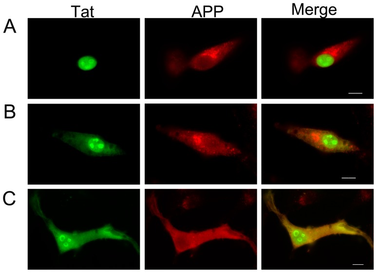
Figure 2. HIV-1 Tat colocalizes with APP in U-87 MG cells.
Fluorescence microscopy images of Tat-transfected U-87 MG cells immunostained with anti-Tat and anti-APP (A8717) antibodies are shown. U-87 MG cells were transfected with the wild-type Tat construct and incubated for 16 h. The cells were fixed and stained with anti-Tat or anti-APP antibodies followed by FITC-conjugated anti-mouse or rhodamine-conjugated anti-rabbit antibodies, respectively. Nuclear (A) and nuclear plus cytosolic (B and C) localization of Tat is shown. Scale bar = 10 µm.

