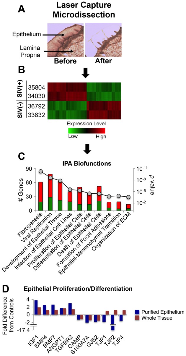
Figure 10. Transcriptional profiling of the macaque tongue epithelium.

A) Representative images of rhesus macaque tongue tissue sections “before” and “after” laser capture microdissection of the epithelial layer. B) Hierarchical clustering of genes up- and down-regulated specifically in the tongue epithelium of SIV infected macaques (35804, 34030) in comparison to uninfected controls (36792, 33832). C) IPA©-based pathway analysis of genes up- and down-modulated in the tongue epithelium of SIV infected macaques. Statistically over-represented biological functions were identified in the list of differentially expressed genes in the tongue epithelium and are shown as stacked bar graphs with the contribution (# genes) from up-regulated transcripts in red and down-regulated transcripts in green. D) Comparison of fold changes in genes associated with epithelial proliferation and differentiation identified through analysis of epithelium purified by laser capture microdissection versus analysis of whole lingual tissues.
