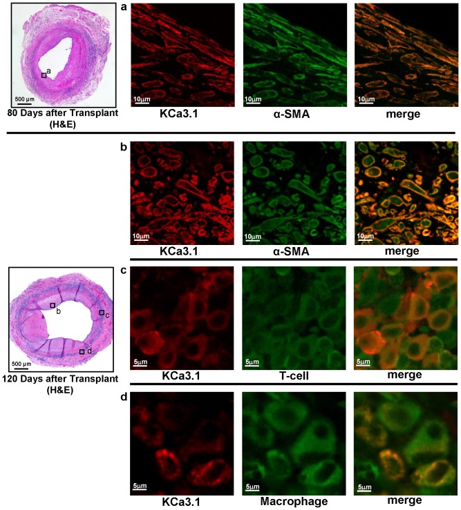
Figure 2. KCa3.1 staining localizes to smooth muscle cells, T cells and macrophages.
Double fluorescent immunostaining for KCa3.1 and α-SMA, T cells (CD43) and macrophages (ED1, CD68) in orthotopic rat aorta transplants harvested 80 or 120 days after transplantation. The boxed areas in the H&E stained vessels on the right show the location where the fluorescent images were taken.

