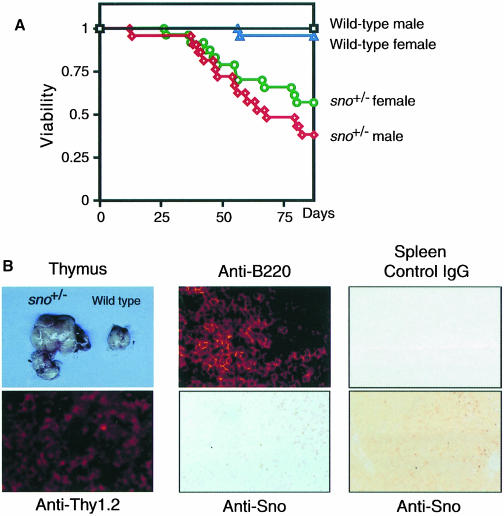
Fig. 4. Chemical carcinogen-induced tumor formation in sno+/– mice. (A) Survival curves of DMBA-treated wild-type and sno+/– mice. For all informative cases, the cause of death was the development of a clinically apparent and histologically confirmed tumor. (B) Histopathological analysis of tumors developed in DMBA-treated sno+/– mice. Left panels: T lymphoma. The enlarged thymus of a tumor-bearing sno+/– mouse is shown (above). The tumor was positive for the T-cell marker Thy-1.2 (below). Center panels: B lymphoma in lymph node. The tumor was positive for B220 (above). Immunostaining with anti-Sno antibody (below). Note that nuclei of normal lymphocytes were stained with anti-Sno antibody, but those of tumor cells were not. Right panels: germinal center in wild-type spleen as a control. Staining with control IgG (above) or anti-Sno antibody (below).

An official website of the United States government
Here's how you know
Official websites use .gov
A
.gov website belongs to an official
government organization in the United States.
Secure .gov websites use HTTPS
A lock (
) or https:// means you've safely
connected to the .gov website. Share sensitive
information only on official, secure websites.
