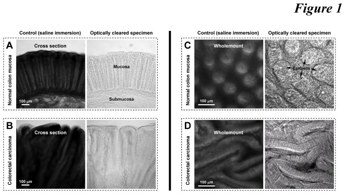
Figure 1. Optical clearing of human colorectal biopsies.
(A and B) Cross sections of normal mucosa and colorectal carcinoma. (C and D) Wholemounts of normal mucosa and colorectal carcinoma. Arrows in panel C indicate the honeycomb-like crypt structure. In panel D, the honeycomb-like crypt epithelia changed to extended layers of epithelia in colorectal carcinoma. Both normal and diseased colons were sectioned by vibratome to specimens of 300 μm in thickness prior to saline or clearing immersion.

