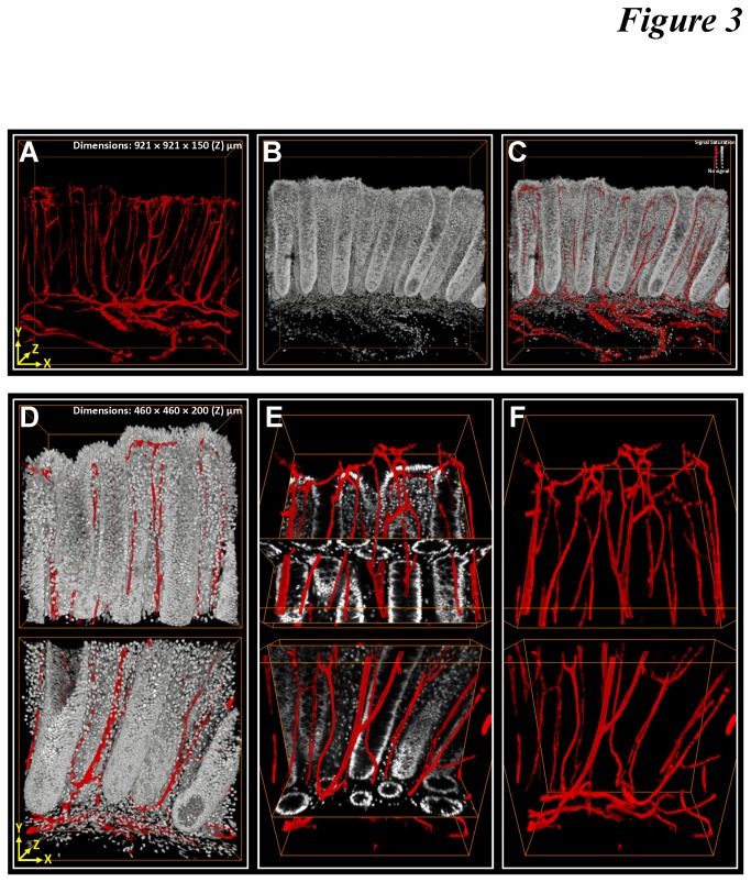
Figure 3. 3-D image rendering and projection of colonic microstructure and vasculature.
(A-C) Gross views of individual and merged projections of colonic crypts (gray: nuclei) and their surrounding vasculature (red: CD34). (D-F) Zoom-in examination of upper and lower parts of the crypts. In panel E, merged projection of vasculature with the orthogonal view of the crypt structure allows the interior domain of the scanned volume to be examined.

