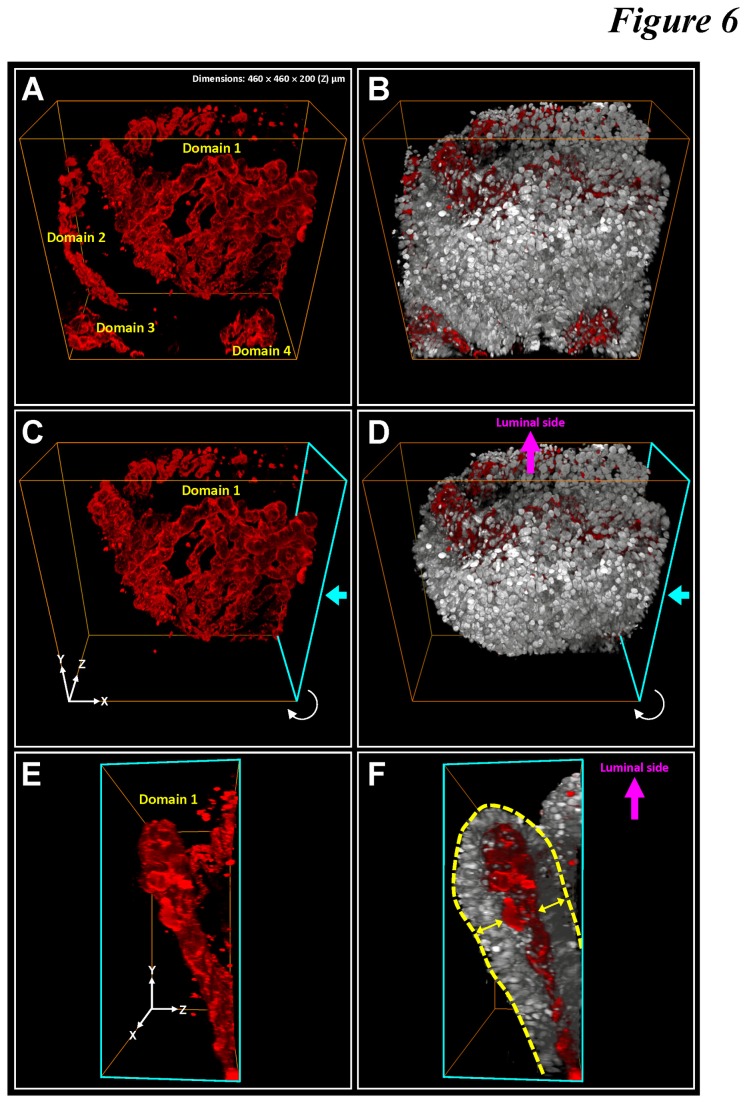
Figure 6. Layer-like microvascular network folded in space in adenocarcinoma.
(A and B) 3-D projection of tumor microstructure (gray: nuclei) and vasculature (red: CD34). Four vascular domains were labeled in the image stack. Prominent nuclear signals were seen around the microvessels. (C-F) Layer-like vasculature folded in space and attached with perivascular cuffs of tumor cells. The signals were segmented from Domain 1 in panel A. Two viewing angles are presented here. The cyan arrows in panels C and D indicate the front side of the projections shown in panels E and F. The yellow arrows in panel F indicate the perivascular cuffs of tumor cells, forming a tumor cell-capillary-tumor cell sandwich structure folded in space (panels C and D). Panoramic projection of the image stack and an additional example of the layer-like microvascular network are presented in Video S4.

