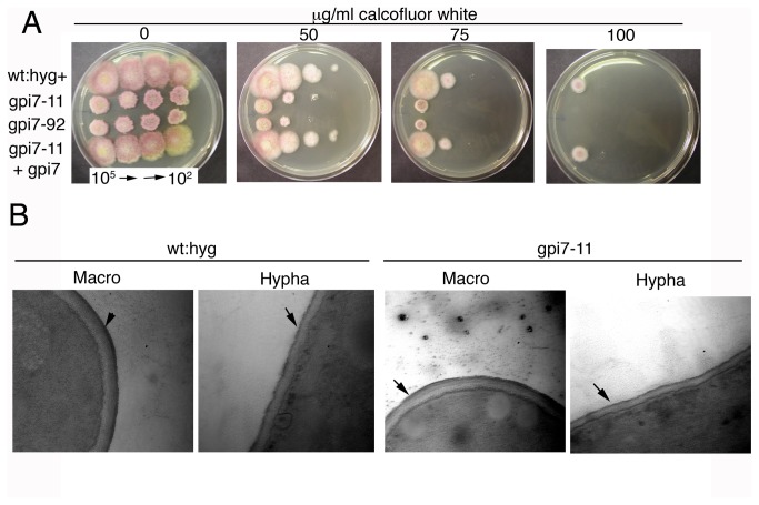
Figure 4. Cell wall defects of Δgpi7 mutants.
A. Increased sensitivity of Δgpi7 mutants gpi7-11 and gpi7-92 to the cell wall disturbing agent calcofluor white compared to the complemented strain (gpi7-11 + gpi7) and hygromycin-resistant PH-1 strain (wt:hyg+). 7 μl of macroconidial suspensions of different concentration were serially spotted onto media. B. Transmission electron micrographs of cell walls in wildtype and Δgpi7 mutant macroconidia and hyphae. Black arrows point to the outer protein layer of the cell wall. There were no gross morphological changes observed in the cell wall of the Δgpi7 mutant. Scale bar = 500 nm.

