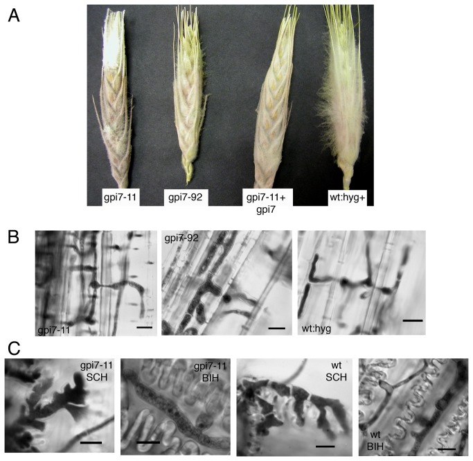
Figure 7. Saprophytic growth of Δgpi7 mutants and development of infection-related hyphae.
A. Previously-frozen wheat heads that were inoculated at a single spikelet and observed 5 days post inoculation. Note the white aerial mycelia profilerating on wheat heads. B. Hyphal development within the rachis tissue of wheat heads depicted in panel A. Micrographs in panel B show the parenchyma cell adjacent to vascular tissue, which were not readily invaded by Δgpi7 mutants in living wheat plants. C. Development of infection-related hyphae on detached wheat glumes. SCH=subcuticular hyphae. BIH=bulbous infection hyphae. Scale bar = 10 μm in all micrographs.

