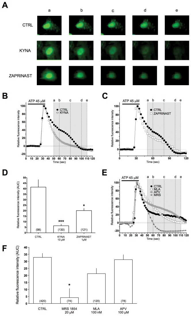
Figure 2. KYNA and zaprinast decrease the [Ca2+]i plateau phase following an IP3-mediated stimulus in mouse cortical astrocytes.
A) A typical example of KYNA and zaprinast-induced decrease in somatic fluo-3 fluorescence time course measured in three different astrocytes in cultures: control (upper panels), in the presence of 10 μM KYNA (middle panels) or zaprinast 1 µM (lower panels). B and C) Time course of KYNA and zaprinast effects on fluo-3 fluorescence in a single astrocyte in culture. KYNA and zaprinast were applied 5 min before the recording was started. D) Area under the curve (AUC) of fluorescence intensity plot following an IP3 induced stimulus in control, KYNA and zaprinast, respectively. E) Time course of MRS-1845, D-APV (50 µM) and MLA (100 nM) effects on fluo-3 fluorescence in a single astrocyte in cultures. F) AUC following an IP3-induced stimulus in control, MRS-1845, D-APV and MLA. Area was calculated from 20 to 70 seconds after the ATP-induced fluorescence peak.

