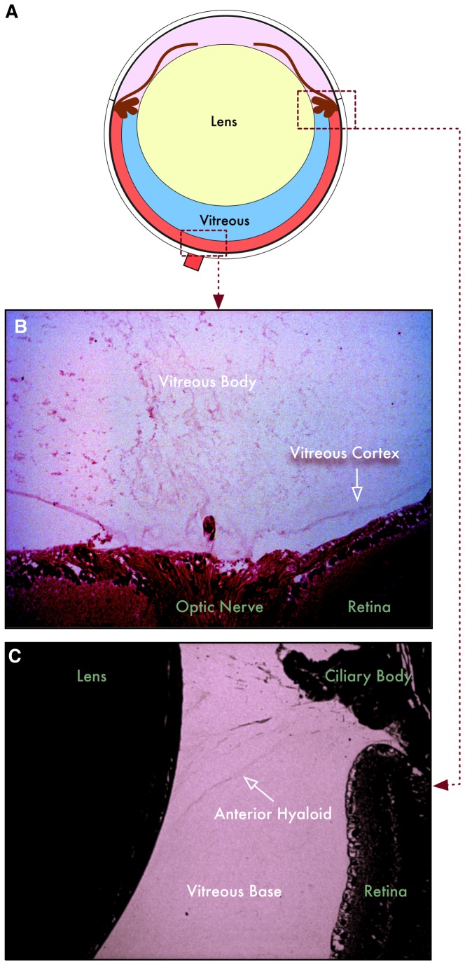
Figure 1. Structure of mouse vitreous.
A. Illustration of the mouse eyeball. The lens composes a large portion of the eye, leaving a small portion of the eye to be filled with vitreous. B. The posterior mouse vitreous has a cortex and body similar to the human vitreous. The cortex defines the vitreoretinal boundary in both human and mouse. C. The mouse anterior hyaloid lies between the ciliary body and lens, posterior to the zonules, anterior to the vitreous base.

