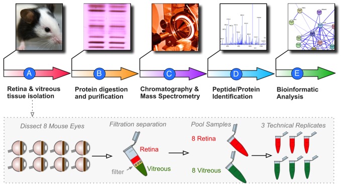
Figure 2. Proteomic analysis pipeline.
A. The vitreous and retina were dissected from 16 mouse eyes. Each tissue type was pooled. B. Protein fractions were isolated and digested with trypsin. C. Peptide fragments were analyzed by multi-dimensional LC-MS/MS. D. MASCOT and SEAQUEST were used for peptide fragmentation finger-printing. E. Proteins with at least 2 peptide hits were analyzed for differential expression, ontology, pathway representation, and protein interactions.

