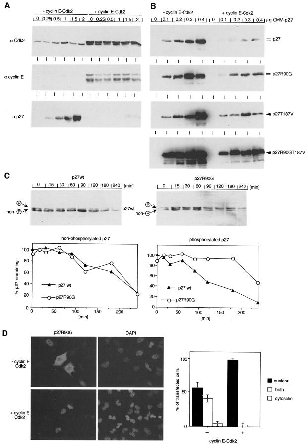
Fig. 6. Cyclin E–Cdk2-induced degradation requires interaction of p27 with mNPAP60. HeLa cells were transfected with the indicated expression plasmids and analysed by Western blotting. (A) Control experiment documenting expression of cyclin E, Cdk2 and p27wt after transfection of the indicated amounts of plasmid; 5 µg of CMV–cyclin E and CMV–Cdk2 were used. (B) Repeat of the experiment using the indicated alleles of p27. Double lines indicate the presence of a phosphorylated form of p27; single arrows indicate absence of phosphorylated p27. (C) Cycloheximide-chase experiment documenting enhanced stability of phosphorylated p27R90G relative to p27wt. Transfections were carried out as in (B). The lower panels show a quantitation of the results and the upper panels show Western blots from a representative experiment. (D) p27R90G accumulates in the nucleus upon expression of cyclin E and Cdk2. Shown are immunofluorescence pictures of HeLa cells transfected with the expression vectors indicated together with a quantitation of the experiment.

An official website of the United States government
Here's how you know
Official websites use .gov
A
.gov website belongs to an official
government organization in the United States.
Secure .gov websites use HTTPS
A lock (
) or https:// means you've safely
connected to the .gov website. Share sensitive
information only on official, secure websites.
