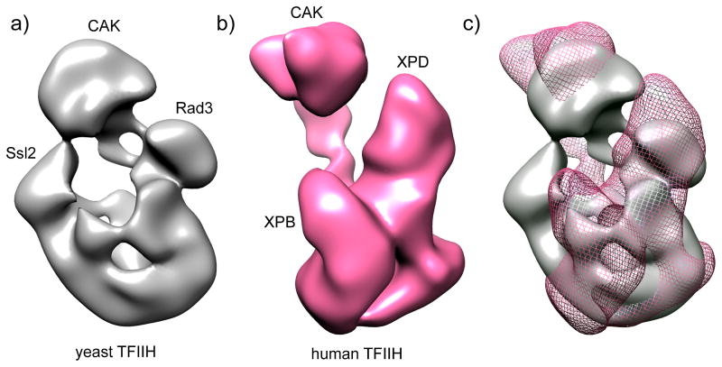
Fig. 2. Structure of TFIIH.
The modules containing the cyclin-dependent kinase (CDK)-activating kinase complex (CAK), Ssl2/XPB, and Rad3/XPD are labeled.
a) Negative stain EM image of yeast TFIIH [41]. The locations of subunits Ssl2, Rad3 and the CAK module (identified by EM analysis of TFIIH lacking these subunits or by gold labeling) are indicated.
b) Negative stain EM image of human TFIIH [21].
c) Alignment of both densities, showing yTFIIH in grey and hTFIIH as pink mesh. EM data suggest a similar general TFIIH shape with several significant differences.

