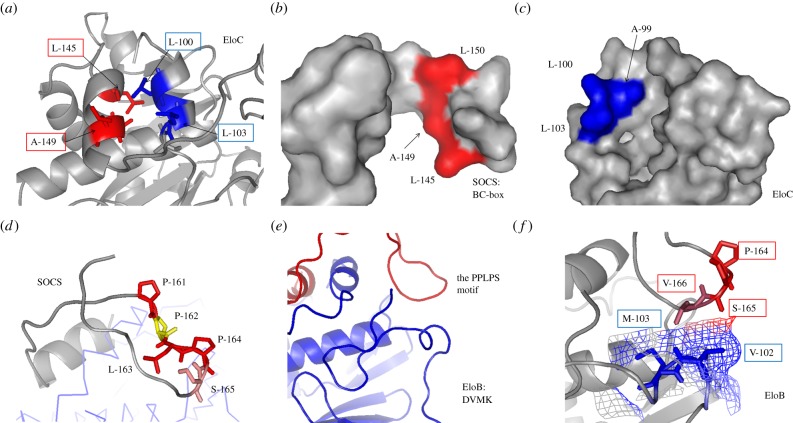
Figure 5.
Interaction between the SOCS-box and EloBC. (a) The interface of the BC-box and EloC binding site. Active side-chains are presented and labelled. (b) The binding surface of BC-box. Resides are coloured in red. (c) The binding surface of EloC C-terminal α-helix. Resides on the interface are coloured in blue. (d) The proline-rich motif downstream from the SOCS-box is coloured. Residues are highlighted in various colours. The second proline (residue 162) in yellow is buried in the complex. (e) The C-terminus of EloB, including the DVMK stretch. Blue, EloB; red, SOCS Peptide. (f) The interface of EloB DVMK stretch and the Vif proline-rich motif. The van der Waals radii of V102 and M103 are presented. The interacted regions by the PPLPS motif are coloured differently.

