
Figure 1.
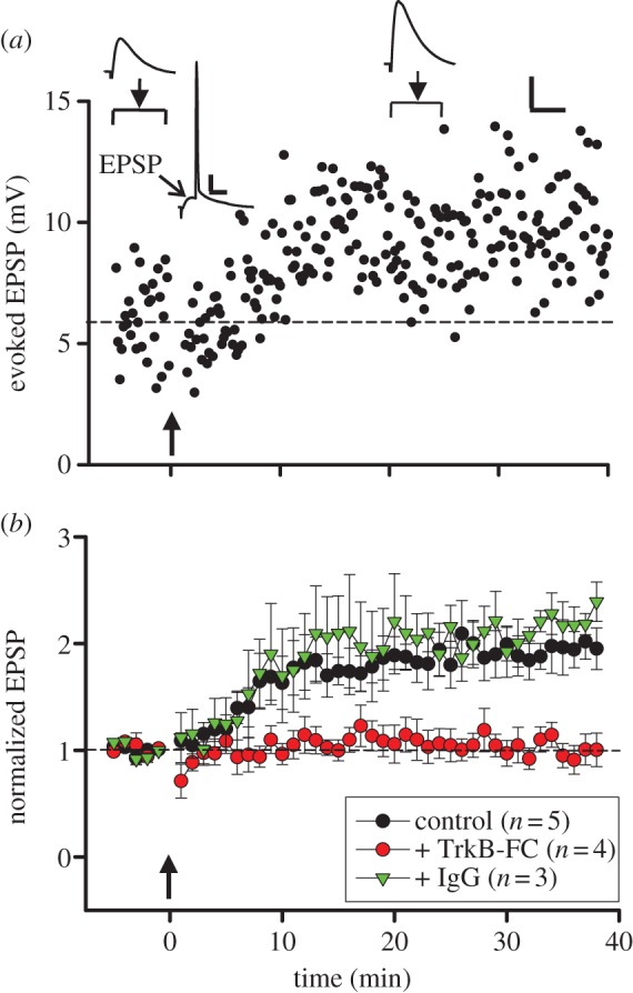
LTP induction in rat hippocampus depends on extracellular BDNF. (a) Example data on changes in the EPSP amplitude induced by paired spiking of pre- and postsynaptic neurons (pre-before-post, Δt = 20 ms, arrow) recorded in hippocampal CA1 pyramidal cells in the brain slice from a rat at P18. Sample traces above: averages of 30 EPSPs (arrowhead) and averaged trace of 80 neuronal responses triggered by paired spiking (Δt = 20 ms). Arrowhead: peak of EPSP. Scales: 4 mV, 50 ms for averaged EPSCs and 10 mV, 20 ms for averaged trace of neuronal responses. (b) Summary of all experiments similar to those in (a), showing EPSP amplitude (mean ± s.e.m., normalized by the mean baseline amplitude) before and after paired pre- and postsynaptic spiking (Δt = 20 ms, arrow) in control artificial cerebrospinal fluid (aCSF; black circle) or in the presence of TrkB–Fc (5 μg ml−1, red circle) or IgG (5 μg ml−1, green triangle).