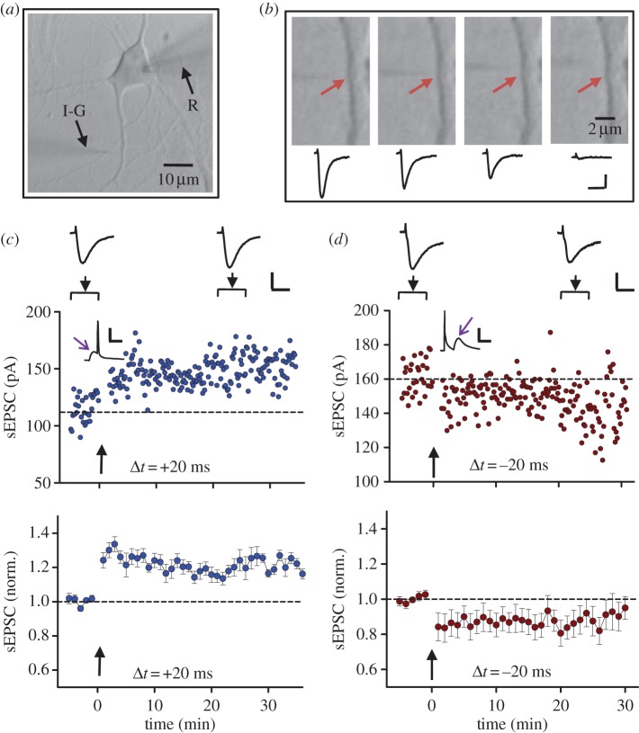
Figure 2.
Glutamate iontophoresis and spiking-timing-dependent potentiation and depression of glutamate responses. (a) Bright field image of a days in vitro 14 (DIV 14) cultured hippocampal neuron. R, recording electrode; I-G, puffing microelectrode for iontophoresis of glutamate. (b) Dependence of response on the position of pipette tip relative to a spine. The response was evoked by iontophoretic application of 150 mM glutamate (pH 8), from a micropipette with a tip less than 1 μm (approx. 200 mΩ). The corresponding current traces include iontophoretic stimulus artefacts (small outward-going pulses) and inward currents mediated by glutamate receptors. Iontophoretic pulse was −150 nA, 0.6 ms. Arrows indicate the spine. Maximal response was obtained when the tip of the puffing microelectrode was pointed to the spine. The response decreased greatly when the pipette tip was moved along the dendrite to 2 μm away from the spine. (c–d) An example (top) and averaged results (bottom) of LTP-like potentiation (c) and LTD-like depression (d) of glutamate-induced responses, induced by paired iontophoretic glutamate and postsynaptic spikes at intervals of 20 ms (c) and −20 ms (d), respectively. Arrows indicate the onset of paired stimulation. (Online version in colour.)

