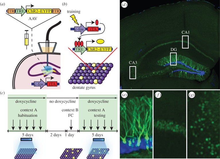
Figure 2.
Basic experimental protocols and selective labelling of the DG cells by ChR2–EYFP. (a) The c-fos–tTA mouse was injected with AAV9-TRE-ChR2-EYFP and implanted with an optical fibre targeting the DG. (b) When off Dox, training induces the expression of c-fos–tTA, which binds to TRE and drives the expression of ChR2–EYFP, labelling a subpopulation of activated cells (yellow) in the DG. (c) Basic experimental scheme. Mice were habituated in context A with light stimulation while on Dox for 5 days, then taken off Dox for 2 days and fear conditioned (FC) in context B. Mice were put back on Dox and tested for 5 days in context A with light stimulation. (d) Representative image showing the expression of ChR2–EYFP in a mouse that was taken off Dox for 2 days and underwent fear conditioning training. An image of each rectangular area in (d) is magnified showing (e) DG, (f) CA1 and (g) CA3. The green signal from ChR2–EYFP in the DG spreads throughout granule cells, including dendrites (e), while the green signal confined to the nuclei in CA1 and CA3 is owing to shEGFP expression from the c-fos–shEGFP construct of the transgenic mouse (f,g). Blue is the nuclear marker DAPI.

