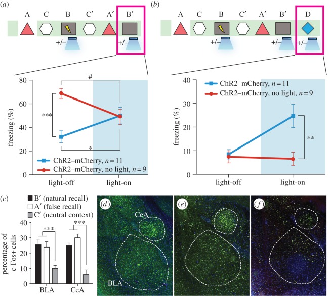
Figure 5.
The false- and genuine-fear memories interact with each other and both recruit the amygdala. (a) Animals that underwent the behavioural protocol shown in figure 2g were re-exposed to context B and the freezing levels were examined both in the absence and presence of light stimulation (n = 11 for ChR2–mCherry group and n = 9 for ChR2–mCherry, no light group; *p = 0.027; ***p < 0.001; #p = 0.034, two-way ANOVA followed by Bonferroni posthoc test). (b) Animals that underwent the behavioural protocol shown in (a) were placed in a novel context D and the freezing levels were examined both in the absence and presence of light stimulation (n = 11 for ChR2–mCherry group and n = 9 for ChR2–mCherry, no light group; **p = 0.007, two-way ANOVA followed by Bonferroni posthoc test). (c) Three groups of mice underwent the training shown in (a) and were sacrificed after testing in either context B (natural recall), A (false recall) or C (neutral context). The percentage of c-Fos-positive cells was calculated for each group in BLA and CeA (n = 6 subjects each; ***p < 0.001). Representative images for natural recall, false recall or neutral context are shown in (d), (e) and (f), respectively.

