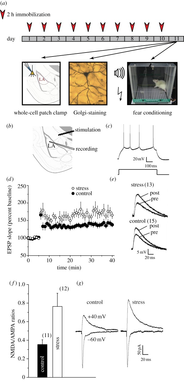
Figure 1.
Chronic stress enhances long-term potentiation (LTP) at thalamic inputs to projection neurons in the lateral amygdala (LA). (a) Schematic of experimental protocol: animals were subjected to 2 h of immobilization, for 10 consecutive days. On the 11th day, animals either underwent fear conditioning, or were sacrificed for Golgi staining or for slice electrophysiology. (b) Placement of recording and stimulating electrodes in a coronal slice of the amygdala. (c) Current-clamp recordings of accommodating action potential firing (top) from a typical LA principal neuron in response to depolarizing current injection (bottom, 600 ms, 0.1 nA). (d) Significantly (*p < 0.05, Student's t-test) greater LTP was induced in stress neurons (open circles, n = 13 cells, from 13 rats) compared with control neurons (filled circles, n = 15 cells, from 15 rats). Summary graphs depict the time course of LTP induced by 30 Hz tetanus (100 pulses per train at 30 Hz, two trains delivered 10 s apart). (e) Superimposed sample traces, from control (bottom) and stress (top) neurons, showing individual EPSPs immediately before (pre) and 35 min after (post) induction of LTP. (f) Summary graph showing significant increase in mean NMDAR/AMPAR-EPSC ratio values in stress neurons (n = 10 cells, 10 rats) compared with controls (n = 11 cells, 11 rats). (g) Traces (averages of 10 recorded responses) of sample AMPAR (−60 mV) and NMDAR (+40 mV) EPSCs from control (left) and stress (right) neurons; traces were selected to obtain matching AMPAR-EPSCs at −60 mV. Error bars are±s.e.m. (Online version in colour.)

