
Figure 1.
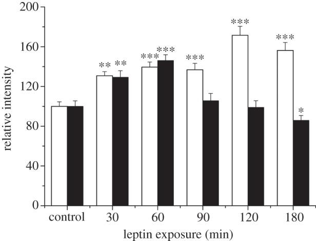
Differential regulation of GluA1 and GluA2 surface expression by leptin. Histogram of pooled data illustrating the effects of leptin on the surface expression of GluA1 (open bars) and GluA2 (filled bars) on hippocampal neurons (7–11 DIC). Leptin (50 nM) evoked a significant increase in GluA1 surface immunostaining after all exposure times (30–180 min). By contrast, exposure to leptin for up to 60 min increased GluA2 surface staining, whereas a significant reduction in GluA2 surface staining was observed after treatment with leptin for between 90 and 180 min.