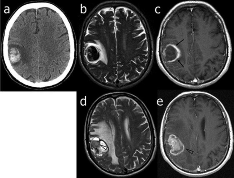
Fig. 1.
Patient 1. Neuroimaging at diagnosis and follow-up. On admission, CT (a), T2-weighted (b) and gadolinium-enhanced T1-weighted (c) axial MR images showed an early, subacute, frontal hematoma with a different wall thickness (arrows) of uncertain nature as well as a vasogenic edema. Two months later, T2-weighted (d) and gadolinium-enhanced T1-weighted (e) MR axial images showed only a slight reduction of the hematoma, an increase of the vasogenic edema and a clearcut evidence of gadolinium-enhanced pathologic solid tissue (arrowheads) consistent with GBM.

