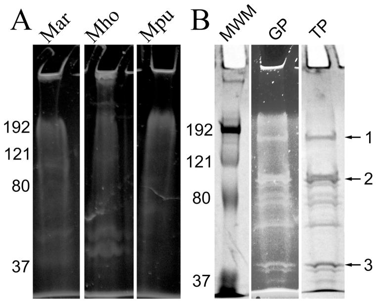
Fig. 1.
Glycoprotein staining of SDS-PAGE gels. Panel A, total proteins from M. arthritidis (Mar), M. hominis (Mho), and M. pulmonis (Mpu), stained for glycoproteins with Pro-Q Emerald 300. Panel B: lane MWM, molecular weight markers (Bio-Rad Kaleidoscope); lane GP, glycoproteins extracted from M. arthritidis with TX-114 and stained with Pro-Q Emerald 300; lane TP, total proteins identified by subsequent staining with Coomassie of lane GP. Numbers on the left of the gels refer to molecular weight standards in kDa. Arrows on the right refer to bands excised for FT-MS analysis.

