
Fig. 2. Phosphorylated tau expression in aged primate brains with developmental exposure to Pb.
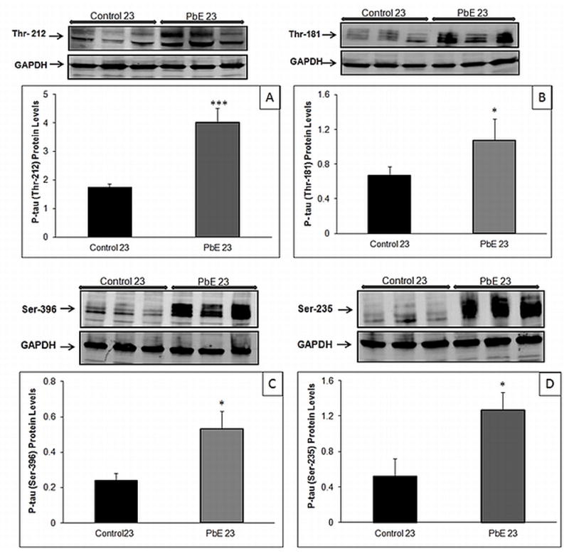
Changes in tau phosphorylation in the cerebral cortex of control and Pb-exposed primate brains (PbE, exposed as infants only) were monitored using Western blot analysis. Relative phosphorylated tau levels (A) Thr-212 (B) Thr-181 (C) Ser-396 (D) Ser-235. Diagrams represents mean +/− SEMs (n=3). Western blot results were normalized against GAPDH. *p<0.05, ***p<0.001 as compared to control 23-year olds.