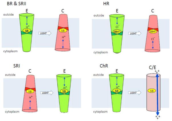
Figure 1. Microbial rhodopsin conformers.
The figure depicts light-induced conformer transitions in the indicated microbial rhodopsins in their native functional state. BR, bacteriorhodopsin; HR, halorhodopsin; SRI, sensory rhodopsin I; SRII, sensory rhodopsin II; ChR, channelrhodopsin. E (green), conformer with externally-connected Schiff base and exterior half-channelopen; C (red), conformer with cytoplasmically-connected Schiff base and cytoplasmic half-channel open; C/E (purple), conformer with an open channel from the extracellular to cytoplasmic surfaces of the protein.

