Figure 1.

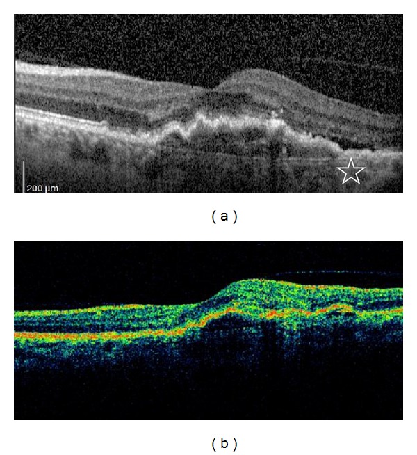
Spectral-domain optical coherence tomography (a) and time-domain optical coherence tomography (b) scans of a patient; the white star shows the subretinal fluid on spectral-domain optical coherence tomography.

Spectral-domain optical coherence tomography (a) and time-domain optical coherence tomography (b) scans of a patient; the white star shows the subretinal fluid on spectral-domain optical coherence tomography.