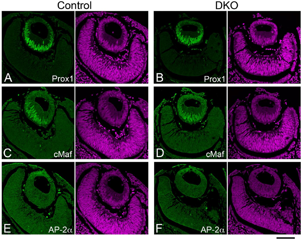
Fig. 4. Induction of early lens markers in Sfrp1;Sfrp2 DKO lenses (E11.5).
Confocal images of paraffin sections immunostained with antibodies indicated in the panels (green). Each panel was paired with PI counterstaining to show nuclei (purple). (A, B) Prox1 is induced in the lens placode and at the lens vesicle stage Prox1 is detected in the elongating primary fibers (A). A normal pattern of expression of Prox1 is maintained in Sfrp1;Sfrp2 DKO lenses (B). (C, D) cMaf is another lens marker with its expression induced in the lens placode and which is subsequently detected in the anterior lens epithelial cells as well as being significantly increased in elongating fibers of the lens vesicle (C). cMaf expression is also maintained in Sfrp1;Sfrp2 DKO lenses (F). (E, F) AP-2α is detected in the anterior lens cells in both control (E) and DKO lenses (F). Scale bar 100µm.

