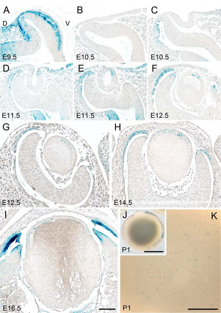
Fig. 5. TCF/Lef reporter activity during lens formation (E9.5-P1).
Coronal paraffin sections of TCF/Lef-lacZ transgenic embryos were reacted with X-gal to detect β-gal activity (A-I). Dorsal to left and ventral to right in all images. (A) At E9.5, strong β-gal activity is detected in neural crest-derived mescenchymal cells between surface ectoderm and optic vesicle but signal is absent in presumptive lens region. (B, C) At E10.5, β-gal reactivity is still not evident in lens placode (B) and lens pit (C). At this stage, X-gal staining is evident in the outer layer of the optic cup on its dorsal side. (D, E) At E11.5, two slices from a same eye through the centre of the lens vesicle (D) and slightly off-centre where lens vesicle is still fused with surface ectoderm (E) are shown. At the neck of lens pit/vesicle blue β-gal activity is asymmetric, being predominantly present on the ventral side. (F) At early E12.5, β-gal activity is present centrally in the anterior layer of the lens vesicle. In the optic cup, X-gal staining is detected at the distal half of the outer layer and the distal tip of the inner layer (presumptive neural retina). (G, H) At late E12.5 and E14.5, X-gal staining is present in the central lens epithelial cells, distal tips of neural retina and distal portion of RPE. (I) At E16.5, X-gal staining in the central lens epithelial cells becomes weaker but is clearly stronger at the distal tips of the optic cup where the ciliary body and iris forms. (J, K) At P1, the whole lens viewed at high magnification shows that cells with β-gal activity are scattered throughout the epithelium. Scale bars for A-I 100 µm, J 1 µm, K 100 µm.

