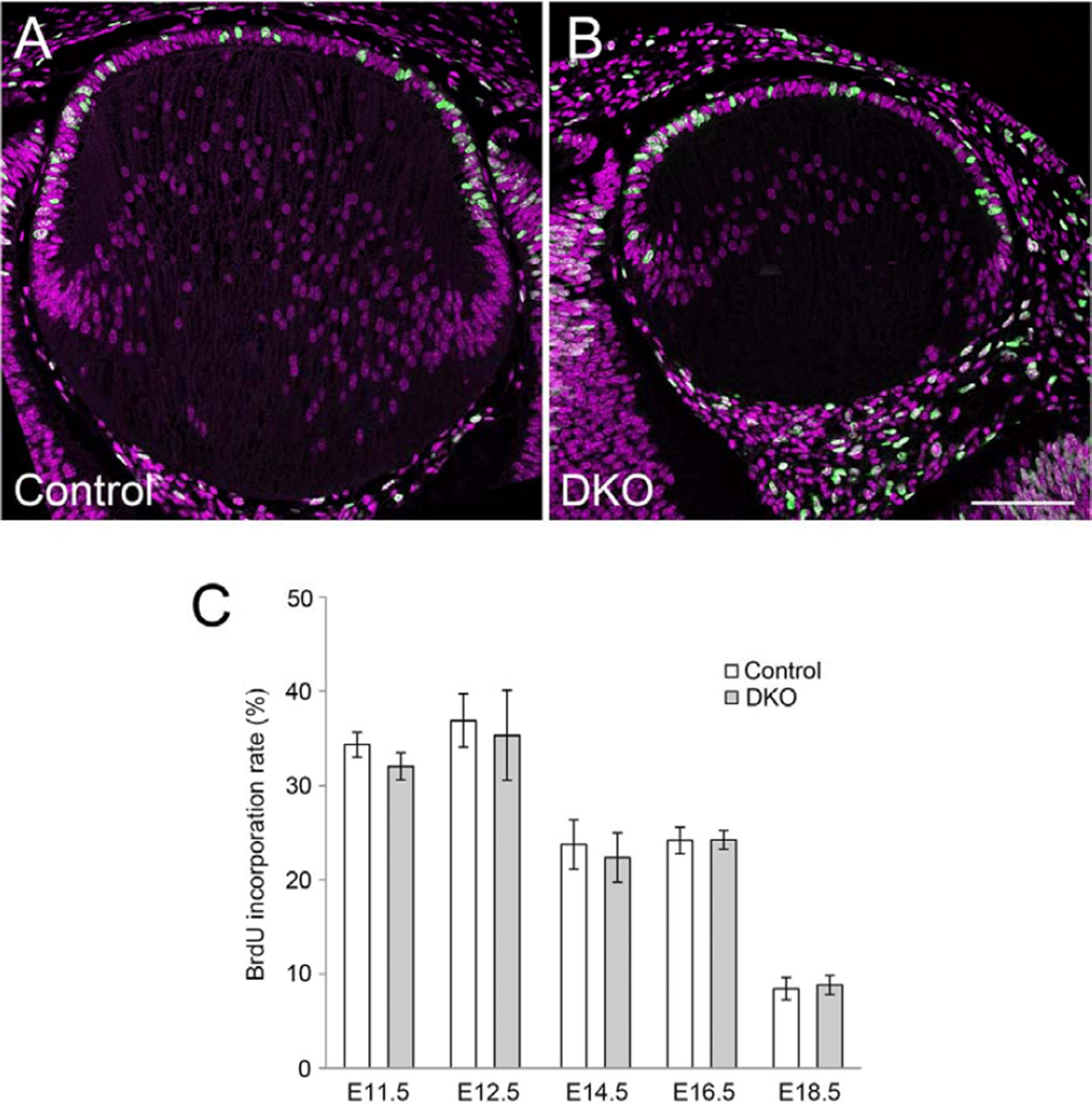
Fig. 7. BrdU incorporation rate does not change in Sfrp1;Sfrp2 DKO lenses.
Pregnant mice were injected with BrdU (100 µg/g body weight) 1 hour before collection of embryos. Incorporated BrdU was visualised by immunostaining with anti-BrdU antibody. (A, B) Confocal images of incorporated BrdU (green) and counterstained PI (purple) in the E14.5 lens. BrdU positive cells and total cell numbers were counted in the central epithelium. Scale bar 200 µm. (C) Mean value of incorporated BrdU (%) in lens cells of lens pit (E11.5), lens epithelial cells located anterior to lens lumen/lens fibers (E12.5) and central lens epithelial cells (E14.5, E16.5 and E18.5). Statistical analysis (two-tailed t-test, α=0.001) does not show any significant differences between controls and DKOs at all ages. Sample number (n) and cells counted were as follows; E11.5 control 1,054 (6), DKO 1,207 (8), E12.5 control 4,748 (18), DKO 2,140 (10), E14.5 control 1,814 (8), DKO 720 (4), E16.5 control 1,617 (6), DKO 1,103 (4), E18.5 control 2, 347 (10), DKO 1,534 (7). Scale bar 200 µm.

